How a Bull Terrier’s Kidney Ultrasound Is Read
Finding out your beloved Bull Terrier needs a kidney ultrasound can be incredibly stressful. You're probably picturing worst-case scenarios and feeling overwhelmed by technical jargon and the fear of the unknown. This comprehensive guide is here to walk you through exactly what a Bull Terrier kidney ultrasound entails, what your veterinarian is looking for, and how those findings are interpreted so you can feel more informed and prepared.
Why Your Vet Recommends a Kidney Ultrasound for Your Bull Terrier
It's natural to feel a knot in your stomach when your vet suggests an advanced diagnostic like an ultrasound. Often, this recommendation comes after routine bloodwork or a urine test, which might have flagged something unusual. Bull Terriers, unfortunately, can be predisposed to certain kidney conditions, making regular monitoring and sometimes more in-depth investigation crucial. Your vet isn't trying to alarm you, but rather to get the clearest possible picture of your Bull Terrier's kidney health.
One of the most common reasons for an ultrasound is an elevated urine protein-to-creatinine (UPC) ratio. If you've been monitoring your Bull Terrier's health, you know that What a UPC Ratio Tells a Bull Terrier Owner is incredibly important. A persistent high UPC ratio, especially in a young or middle-aged Bull Terrier, can be a red flag for a condition like Hereditary Nephritis (HN). While a blood test can show if the kidneys are struggling, an ultrasound helps visualize why they might be struggling, looking for structural changes that blood tests alone can't reveal.
Beyond routine screenings, clinical signs can also prompt an ultrasound. Perhaps your Bull Terrier has started drinking more water than usual, urinating more frequently, or even seems a bit off – lethargic, reduced appetite, or experiencing unexplained weight loss. These Signs a Bull Terrier's Kidneys Are Failing are serious and warrant immediate investigation. An ultrasound provides a non-invasive way to check for issues that might be causing these symptoms, helping to pinpoint the problem area before it progresses further.
What the Vet Is Looking For During a Bull Terrier Kidney Ultrasound
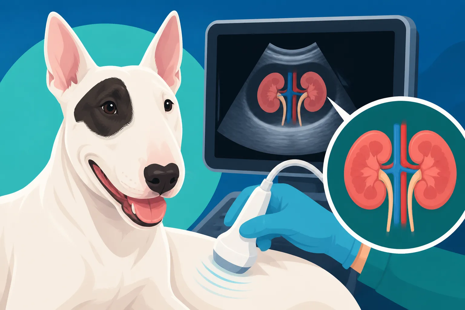
When your Bull Terrier goes in for a kidney ultrasound, it’s not just about getting pretty pictures. The veterinary ultrasonographer, often a specialist, is meticulously examining several key aspects of each kidney. Think of it as a guided tour of the internal architecture of these vital organs, checking for any deviations from what a healthy Bull Terrier kidney should look like. This detailed assessment helps rule out or confirm various conditions, providing critical information for diagnosis and treatment planning.
Assessing Kidney Size, Shape, and Location
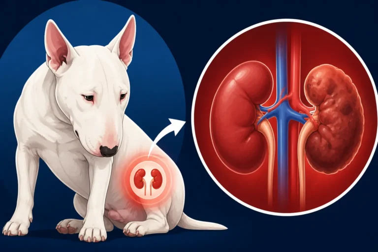
First and foremost, the ultrasonographer will evaluate the size, shape, and exact location of both kidneys. In a healthy Bull Terrier, kidneys are typically bean-shaped and fairly symmetrical, nestled comfortably in the abdominal cavity. Deviations from this norm can be quite telling. For instance, kidneys that are unusually small and shrunken might suggest chronic kidney disease, where the organ has atrophied over time due to long-standing damage. Conversely, an enlarged kidney could point to acute inflammation, infection, or even an obstruction causing fluid buildup, known as hydronephrosis.
The overall contour of the kidney is also important. A smooth, well-defined outline is ideal. Any irregularities, such as bumps or indentations, could indicate a mass, cysts, or scarring from previous injury or disease. Sometimes, the kidneys might be displaced from their usual position, which can happen with large abdominal masses pushing them aside, or in rare cases, due to a congenital anomaly. Understanding these basic morphological characteristics provides the foundational layer for interpreting the more intricate details revealed by the ultrasound.
Evaluating Internal Architecture and Echogenicity
Once the gross anatomy is assessed, the ultrasonographer dives into the internal structure, looking at what's called the "echogenicity" – essentially, how bright or dark different tissues appear on the screen. A normal Bull Terrier kidney has a distinct internal architecture, with clearly differentiated regions: the outer cortex, the middle medulla (which contains the pyramids), and the central renal pelvis. The cortex is usually slightly brighter than the medulla, and the renal pelvis appears as a relatively anechoic (dark) area unless there’s fluid or debris.
Changes in echogenicity and the loss of this clear differentiation between the cortex and medulla are major red flags. If the entire kidney appears uniformly bright, it might suggest diffuse fibrosis or inflammation, which can be seen in conditions like Hereditary Nephritis Takes Bull Terriers in Their Prime. Conversely, abnormally dark areas could indicate fluid accumulation or necrosis. The presence of hyperechoic (very bright) spots might point to mineralization or calcifications, often associated with chronic kidney disease or certain metabolic imbalances. These subtle shifts in the visual texture of the kidney are vital clues for the diagnosing veterinarian.
Checking for Cysts, Calculi, and Masses
Beyond general changes, the ultrasound is excellent for identifying specific abnormalities like cysts, kidney stones (calculi), and masses. Cysts appear as well-defined, fluid-filled, dark (anechoic) structures, and while some small, simple cysts might be benign, multiple or large cysts, especially in certain patterns, can be indicative of conditions like Polycystic Kidney Disease in a Bull Terrier. These are typically congenital and can impair kidney function over time.
Kidney stones, or uroliths, show up as bright, reflective structures within the kidney or renal pelvis, often with an acoustic shadow behind them, meaning the sound waves cannot penetrate beyond the stone. This shadow is a classic sign. They can cause pain, infection, and obstruct urine flow, leading to significant kidney damage. Masses, on the other hand, can be solid or mixed, appearing as irregular areas of differing echogenicity. Distinguishing between benign growths, abscesses, and malignant tumors is critical, and sometimes the ultrasound can provide enough information to guide decisions for further diagnostics, such as a biopsy.
Interpreting the Ultrasound Findings: What the Images Tell Us
The images generated by the ultrasound machine are just one piece of the puzzle. The real magic happens when an experienced veterinary ultrasonographer or radiologist interprets these images in conjunction with your Bull Terrier's clinical signs, bloodwork, and urinalysis results. They are looking for patterns and specific characteristics that point towards a definitive diagnosis. It’s not always black and white, but rather a complex interplay of findings that helps paint a clear picture of what’s going on inside.
Identifying Signs of Hereditary Nephritis
For Bull Terriers, one of the most concerning conditions an ultrasound might reveal is Hereditary Nephritis (HN). While a definitive diagnosis often requires a kidney biopsy, the ultrasound can provide strong supportive evidence. In early stages, kidneys affected by HN might look relatively normal. However, as the disease progresses, the ultrasound often shows signs of increased cortical echogenicity – meaning the outer layer of the kidney appears brighter than it should be, losing the clear distinction between the cortex and the medulla. This indicates diffuse fibrosis and scarring.
As How Hereditary Nephritis Damages a Bull Terrier's Kidneys, the kidneys become progressively damaged. Over time, Bull Terriers with HN might develop kidneys that are smaller than normal, with an irregular outline due to the scarring. The internal architecture becomes indistinct, almost a uniform bright gray, indicating severe and widespread disease. The presence of subtle changes, even in the absence of significant kidney failure on blood tests, can prompt a veterinarian to recommend further tests, such as genetic screening or a biopsy, to confirm HN and initiate early management strategies.
Detecting Obstructions and Hydronephrosis
One of the significant advantages of an ultrasound is its ability to visualize obstructions within the urinary tract and their consequences. If a kidney stone, tumor, or even a stricture is blocking the flow of urine from the kidney down the ureter to the bladder, the urine can back up and cause the renal pelvis to distend. This condition is called hydronephrosis. On ultrasound, hydronephrosis appears as an enlarged, fluid-filled renal pelvis, often with a thinned renal cortex.
Detecting hydronephrosis is critical because if left untreated, the continuous pressure from the backed-up urine can cause irreversible damage to the kidney tissue. The ultrasound can pinpoint the exact location and nature of the obstruction, whether it's a stone, a clot, or a tumor. This information is invaluable for determining the appropriate intervention, which could range from medical management to surgical removal of the obstruction, to preserve kidney function.
Differentiating Benign from Concerning Growths
When a mass or abnormal growth is identified, the ultrasound helps characterize it to determine if it's benign or potentially malignant. A simple renal cyst, for example, will appear perfectly anechoic (black) with smooth, thin walls and enhanced through-transmission (sound waves pass easily through it). These are usually not a major concern. However, if a mass has irregular borders, a mixed echogenicity (areas of bright and dark), or shows signs of invading surrounding tissue, it raises suspicion for a more aggressive tumor.
The ultrasonographer will also use Doppler flow studies to assess blood flow within and around the mass. Tumors often have aberrant and increased blood supply, which can be visualized with color Doppler. While an ultrasound cannot definitively diagnose cancer – that typically requires a biopsy and histopathology – it provides crucial information about the size, location, and characteristics of the mass, guiding the next diagnostic or therapeutic steps. Understanding these nuanced differences helps pet owners and vets make informed decisions together, ensuring the best possible outcome for their Bull Terrier.
| Kidney Ultrasound Finding | Appearance on Ultrasound | Potential Implication |
|---|---|---|
| Normal Kidney | Bean-shaped, smooth outline, clear cortex/medulla differentiation, moderate echogenicity. | Healthy function, no gross abnormalities. |
| Small, Irregular Kidneys | Reduced size, scarred or bumpy outline, poor cortex/medulla differentiation, often hyperechoic. | Chronic kidney disease, end-stage renal failure, hereditary nephritis. |
| Enlarged Kidneys | Increased size, can be smooth or irregular outline. | Acute kidney injury, inflammation, infection, hydronephrosis, tumor. |
| Increased Cortical Echogenicity | Outer kidney layer appears brighter than normal. | Fibrosis, inflammation, amyloidosis, Hereditary Nephritis. |
| Loss of Medullary/Cortical Definition | Internal layers indistinct, kidney appears uniformly textured. | Severe chronic kidney disease, diffuse inflammation. |
| Cysts | Anechoic (dark), round structures with smooth walls. | Benign cysts, Polycystic Kidney Disease (if multiple). |
| Calculi (Stones) | Hyperechoic (bright) spots with acoustic shadowing. | Urolithiasis, potential obstruction, infection. |
| Masses/Tumors | Irregular shape, mixed echogenicity, abnormal blood flow (on Doppler). | Neoplasia (benign or malignant), abscess, hematoma. |
| Hydronephrosis | Dilated, fluid-filled renal pelvis, thinned cortex. | Urinary obstruction (stone, tumor), ureteral stricture. |
Next Steps After a Bull Terrier Kidney Ultrasound
Receiving the results of your Bull Terrier’s kidney ultrasound can bring a mix of emotions – relief, concern, or sometimes just more questions. What happens next largely depends on what the ultrasound revealed. The good news is that an ultrasound is a powerful diagnostic tool that usually provides a clear direction for managing your Bull Terrier’s health moving forward. It’s important to remember that this isn't the end of the road, but a crucial step in ensuring your furry companion gets the best possible care.
Further Diagnostics and Treatment Planning
If the ultrasound reveals significant abnormalities, your vet will likely discuss additional diagnostic tests. For instance, if a suspicious mass was found, a fine-needle aspirate or a biopsy might be recommended to determine if it's cancerous. This usually involves taking a small tissue sample for laboratory analysis. If hydronephrosis is present, further imaging like a CT scan might be needed to precisely map the obstruction, or a specialist consultation with a veterinary urologist might be advised. Don’t hesitate to ask your vet for clarity on why each test is needed and what it aims to discover.
Based on these findings, a specific treatment plan will be formulated. This could range from medication to manage inflammation or infection, dietary changes to support kidney function, or even surgery in cases of large stones, tumors, or severe obstructions. Sometimes, even if the findings are minor, your vet might recommend close monitoring with regular The Yearly Urine Test Every Bull Terrier Owner Should Book or follow-up ultrasounds to track any progression. Having pet insurance like Healthy Paws can be a real lifesaver during these times, as it helps cover the costs of these specialized diagnostics and treatments, easing the financial burden and allowing you to focus on your Bull Terrier's well-being.
Long-Term Management and Prognosis
For many Bull Terriers, especially those diagnosed with chronic kidney disease or conditions like Hereditary Nephritis, the journey often involves long-term management. This means ongoing dietary modifications, often a prescription renal diet specifically designed to reduce the workload on the kidneys and slow disease progression. You might need to regularly monitor your Bull Terrier's water intake, urine output, and weight. Regular veterinary check-ups, often every three to six months, will become a part of your routine to track kidney values and adjust medications as needed. This proactive approach to Caring for a Bull Terrier Past Eight is crucial.
The prognosis varies widely depending on the specific condition, its severity, and how early it was caught. With conditions like Hereditary Nephritis, while there's no cure, early diagnosis and diligent management can significantly improve your Bull Terrier's quality of life and potentially extend their lifespan. Your veterinary team will be your partner throughout this process, providing guidance, support, and adjusting the care plan as your Bull Terrier's needs evolve. Remember, you’re not alone in this, and focusing on consistent, compassionate care will always be the best path forward.
Disclaimer
This article is intended for informational purposes only and does not constitute veterinary medical advice. Always consult with a qualified veterinarian for any health concerns or before making any decisions about your Bull Terrier's care.